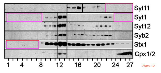
Can the authors please take a look at Figure 1D? I have some concerns.
- Pink boxes: the same area appears to be visible four times: one time in the Syt11 blot, two times in the Syt 1 blot, and one time in the Stx1 blot.
- The area is stretched differently on each occasion
- I have converted the image to black and white before I added my pink boxes, because it has red and blue boxes by itself, which would be confusing.