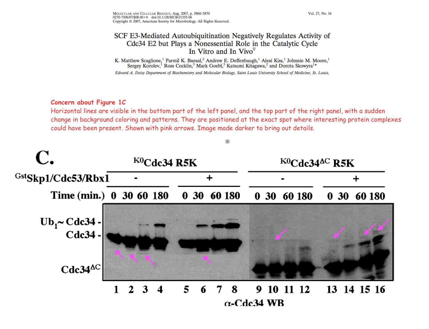
1. Concern about Figure 1C.
Horizontal lines are visible in the bottom part of the left panel, and the top part of the right panel, with a sudden change in background coloring and patterns. They are positioned at the exact spot where interesting protein complexes could have been present.
See my concerns about Figure 1C here:
Concern about Figure 4B and 4C.
In the top panel of Figure 4B-b, a cut appears to be visible between the left and right half of the blot image.
In the bottom panel of Figure 4B-b, a horizontal “patch” appears to be visible, with a lighter background than the rest of the blot.
In Figure 4C, vertical lines are visible.
See my concerns about Figure 4 here: