Copied from: https://pubpeer.com/publications/1D808F3B2ED8155399A41F2D835F6A
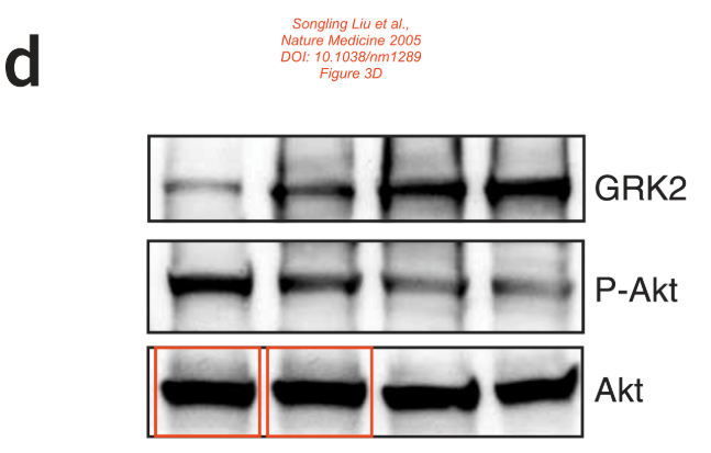
Concern about Figure 3D:
Red boxes: The first two lanes of the Akt panel look unexpectedly similar.
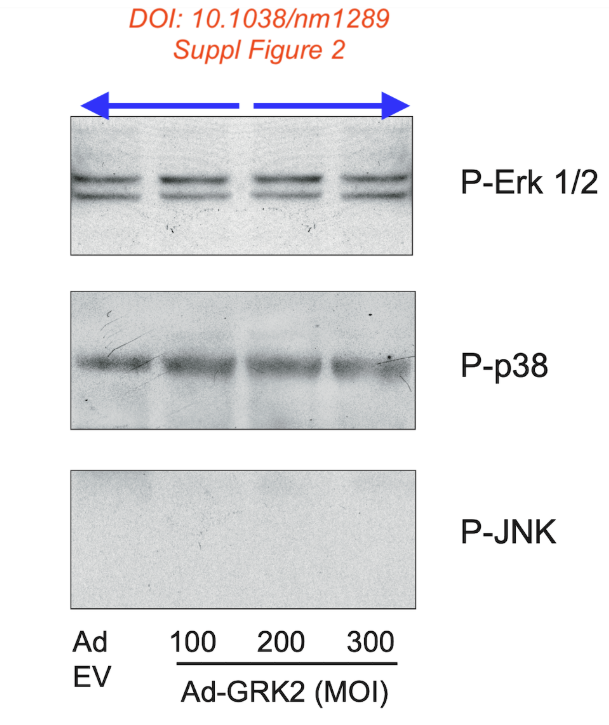
Concern about Supplementary Figure 2.
Blue arrows: p-Erk1/2 blot: Lanes 1 and 2 look remarkably similar to lanes 3 and 4 in mirror image.
As much as I admire symmetry, I would not expect that in this blot. Could the authors provide a satisfactory explanation, please?